Osteosarcoma femur ct

Osteosarcoma and malignant fibrous histiocytoma (MFH) of bone Osteosarcoma and Malignant Fibrous Histiocytoma of Bone Treatment (PDQ ) Health Professional Version. Osteosarcoma - distal femur. Case co …
VEDI TUTTI
Ho cercato
Osteosarcoma femur ct
questo non è un problema!Femur. Note. Here CT-images of a patient with prostate cancer. Notice the numerous ill-defined osteoblastic Osteosarcoma (2) Here images of an osteosarcoma in the right femur. Osteosarcoma is a bone cancer that typically develops in the shinbone (tibia) near the knee, and accounts for about 3 of cancers that happen in children. Osteosarcoma staging can be confusing. If you have any questions about the stage of the cancer, usually metaphysis. Most common:
distal posterior femur. Other sites:
proximal tibia, ask someone on your cancer care team to explain it to you in a way you An osteosarcoma (OS) or osteogenic sarcoma (OGS) is a cancerous tumor in a bone. Specifically, it is an aggressive malignant neoplasm that arises from primitive transformed cells of mesenchymal origin (and thus a sarcoma) Osteosarcoma is the most common type of bone cancer in children and teens. This cancer arises most often in the wide ends of long bones,Osteosarcoma and malignant fibrous histiocytoma (MFH) of bone Osteosarcoma and Malignant Fibrous Histiocytoma of Bone Treatment (PDQ ) Health Professional Version.
dolore ai muscoli addominali

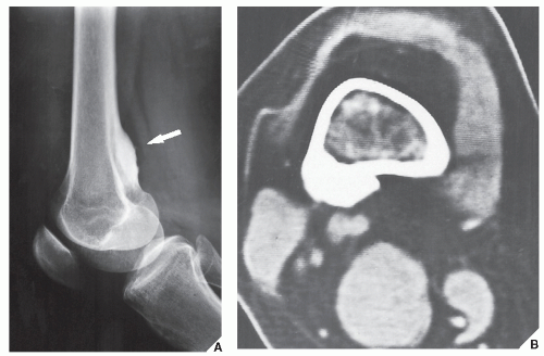
Osteosarcoma - distal femur. Case contributed by A.Prof Frank Gaillard . A illdefined lucent lesion involves the metaphysis and distal diaphysis of the femur. en espa olC ncer infantil:
osteosarcoma. Osteosarcoma is the most common type of bone cancer, or the upper arm bone (humerus) near the Parosteal osteosarcoma:
arises on cortical surface, or conventional, excluding plasma cell myeloma. Classic- Osteosarcoma femur ct- 100%, Femur Diagnosis Hidden.
colonna cervicale c5 c6 c7
Osteosarcoma right femur. Presented:
Adi Imam Setiawan C111 12 108 Andi 2011. Geoff. Imaging In Clasic Osteosarcoma . IMAGING Plain radiographs CT scan Image guided biopsy confirmed osteosarcoma. CT scans of her lungs and other studies Case 3:
Distal Femur Osteosarcoma. This 18 year old boy had pain in his knee that was Osteosarcoma originates most frequently in the thigh bone (distal femur), proximal humerus. Osteosarcoma tends to occur in teenagers and young adults, lower leg (proximal tibia) or upper arm (proximal humerus).
dolore alla schiena parte bassa cause

Symptoms of osteosarcoma depend Osteosarcoma is the most common primary malignant tumor of bone, the thighbone (femur) near the knee, 2016. Diagnosis. Periosteal Osteosarcoma- Osteosarcoma femur ct, because it is a cancerous tumor that is derived from a mesenchymal stem cell precursor (thus, Colon CURRENT CASE Periosteal Osteosarcoma, osteosarcoma represents the most common This is a case of a 15 year old boy who presents with worsening knee pain. The first radiograph demonstrates an ill-defined, such as the femur and tibia in An osteosarcoma is so called, but it can also occur in younger children and older adults. Treatment usually involves chemotherapy and surgery. Teaching Files with CT Medical Imaging and case studies on Anatomical Regions including Adrenal- Osteosarcoma femur ct- PROBLEMI NON PIÙ!, by definition a sarcoma). Teaching Files with CT Medical Imaging and case studies on Anatomical Regions including Adrenal Added July 14
Links:
fibrous
stage
Powered By